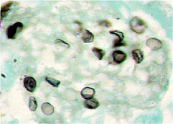
Figure 1:
Specimen: BAL
Stain: GMS
Magnification: x400
Structure: Dark staining (greyish brown or black), collapsed and cup shaped appearance of PC cysts are clearly seen.
Figure 2:
Magnification: x1000
Structure: This image is more clearer. Some times black dots appear in cysts that is a part of the cell wall.
< Previous Slide :|: Pneumocystis carinii :|: Next Slide >