Image Gallery Main Menu Yeasts Opportunistic Fungi Dematiaceous Fungi
Dimorphic Fungi Dermatophytes Actinomycetes Direct Microscopy
Pneumocystis carinii Technical Cases

•Specimen: BAL

•Stain: ZN Stain

•Magnification: x1000


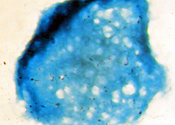
Structure: Most areas in the image of foamy exudate showing honeycomb appearance contain cysts of PC. Clusters of PC cysts that remain unclear due to being masked by the counter stain. ZN is not a proper stain for detecting PC, but the image shown here demand further analysis to confirm PC in the specimen.


< Previous Slide :|: Pneumocystis carinii :|: Next Slide >

© Copyright 1999-2007 Department of Microbiology, Mount Sinai Hospital, Toronto, Canada. All rights reserved.