Image Gallery Main Menu Yeasts Opportunistic Fungi Dematiaceous Fungi
Dimorphic Fungi Dermatophytes Actinomycetes Direct Microscopy
Pneumocystis carinii Technical Cases

Figure 1:

• Specimen: BAL

• Stain: Fungi-Fluor Stain

• Magnification: x400


Figure 2:

• Specimen: BAL

• Stain: Fungi-Fluor Stain

• Magnification: x400


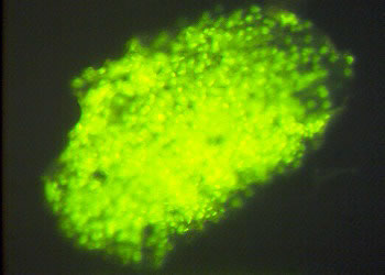
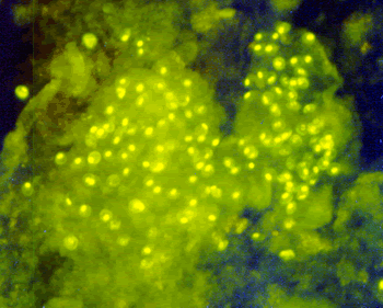
Structure: Fungi-Fluor detects PC nonspecifically. Cysts of PC usually found in clusters embedded in the foamy exudate. PC cysts on the images shown are not so clear since they are crowded and background staining interferes with clarity. As a result they lack the characteristic morphology of the PC cysts.


< Previous Slide :|: Pneumocystis carinii :|: Next Slide >

© Copyright 1999-2007 Department of Microbiology, Mount Sinai Hospital, Toronto, Canada. All rights reserved.