9-year-old Female with Fever and Headache
Presented by:
Dr. D. Yamamura1 and Mr. L. Wilcox2
1MDS Laboratories and 2Hamilton
Regional Laboratory Medicine Program
Clinical Case:
A previously healthy 9-year-old female was admitted to Children's
Hospital with a 10 day history of fever up to 103ºF and headache.
The patient developed a raised red rash with some pustules 7 days
prior to admission beginning on her lower extremities and soles
of her feet progressing to involve her upper extremities, palms,
face and trunk. Multiple joints involving her right hand, left
hand, right elbow, left wrist, left shoulder and bilateral knees
and ankle joints were red and swollen. The polyarthritis was asymmetric
and migratory. The patient denied any history of arthritis. There
was no preceding dairrheal or respiratory illness. The patient
was not sexually active and had not travelled. There was no significant
past medical history. A maternal aunt had rheumatoid arthiritis.
The patient was on Acetaminophen and Ibuprofen. All members of
her family had a recent upper respiratory infection. The patient
had a pet rat.
On physical examination, the patient appeared toxic and listless.
She had temperature of 36.9ºC and had an elevated heart rate.
The respiratory and cardiovascular examination was unremarkable.
Swelling, erythema and decreased ROM were seen in multiple joints.
Pustular lesions were seen on the soles of her feet. There was
no enlargement of her lymph nodes, or lesions in the oral cavity.
Laboratory investigation revealed that the patient had a WBC 8.3,
Platelets 328, ESR 85, CRP 168, and a negative ASOT. Rheumatoid
factor was normal. Joint aspirate of her knee showed 45.5 x 10⁹
nucleated cells/L with 89% neutrophils. One of 2 sets of blood
cultures using the BACTEC 9240 (Bectom Dickson Microbiology Systems)
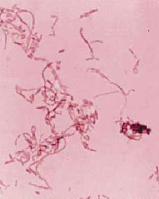
became positive after 28 hours of incubation. The gram stain revealed
a moderate to large, pleomorphic gram negative bacilli (GNB) with
long filaments and irregular swellings (Figure 1). Aspirate of
the right knee did not reveal any organisms by gram stain and
the culture was negative. A pleomorphic GNB was seen on the gram
stain of a swab of a pustule on the right foot.
The patient was treated with intravenous penicillin and gentamicin
and improved clinically. A transthoracic echocardiogram did not
reveal endocarditis. The patient was discharged home on amoxicillin.
Results from further microbiology investigations are summarized.
The blood culture was sub-cultured to sheep blood agar (SBA) incubated
anaerobically, chocolate agar (CA) incubated in 5% CO2, and
MacConkey agar (MAC) with crystal violet incubated aerobically.
No growth was seen on MAC or CA. Pinpoint growth was seen at 48
hours on SBA. The colonies were round, smooth, and gray. Initial
work-up revealed a catalase and oxidase-negative organism. No
reaction was seen on standard biochemical tests. The organism
was incubated in supplemented thioglycollate broth (Figure 2).
Further biochemical tests were performed. A reference laboratory
confirmed the identification.
Questions:
- What are the possible infectious and non-infectious causes
of rash, fever, and polyarthritis? After viewing the gram stain
what are the most likely cause(s)?
- What zoonotic (animal) sources have been linked to this syndrome?
- In a routine microbiology laboratory, what further tests and
supplementation would aid in the diagnosis of this organism?
What reaction do you see with the thioglycollate broth and what
organism is most likely given the reaction? What other diagnostic
modalities are available to confirm the identification?
- What are L-forms and what implication does this have for treatment?
- What clinical complications have been reported with this syndrome?
Figures:
 |
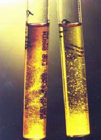 |
Figure 1: Gram stained smear of isolate
on blood agar medium |
Figure 2: Supplemented Thioglycollate
Broth |
Discussion:
This
is a case of Rat-bite Fever caused by Streptobacillus moniliformis.
Rat-bite Fever can also be caused by another gram-negative bacillus,
Spirillum minus. Infection with S. monilformis is
characterized by the sudden onset of fever, sweats, headache and
muscle aches typically followed by polyarthritis and a rash. The
rash can be raised and red, vesicular, or pustular. The distribution
of the rash is an important clinical clue as the extremities,
the soles of feet, and palms are frequently involved. Rarely,
the rash and arthritis are absent. Endocarditis is a rare complication
with increased mortality of up to 50%. Other unusual manifestions
such as genital tract infection, meningitis, amnionitis, pericarditis
have been reported.
Infection caused by S. moniliformis is rare. It is not
a reportable disease so the incidence is unknown. It is a zoonosis
with transmission either by direct contact with a rat (Rat-bite
Fever), usually a bite or scratch, or by ingestion of water or
milk contaminated by rat urine or feces (Haverhill fever). Rarely,
other animals such as guinea pigs, mice, squirrels and gerbils
have been implicated. Colonization of the nasopharynx in wild
rats is reported to be higher than rates in laboratory rats and
the majority of reported cases are due to exposure to wild rats.
Colonization rates in pet rats are not known. This patient had
a pet rat and had also been exposed to her friend’s two
young rats. Although there was no bite the patient did sustain
some scratches from the young rats.
The differential diagnosis of rash and polyarthritis includes
infectious and non-infectious causes. Other important bacterial
causes are N. gonorrhoeae (disseminated gonococcal disease),
N. meningitides, and H. influenzae. Viral pathogens
such as rubella and parvovirus B12 can also cause rash and polyarthritis.
Reiter’s syndrome, which likely occurs due to an immune
response to an infectious agent such as Chlamydia trachomatis
or following bacterial infectious gastroenterititis, can cause
a polyarthritis; however, it is unusual to have a raised red or
pustular rash. Rheumatoid arthritis is the non-infectious syndrome
most likely to be misdiagnosed.
S.
moniliformis is a highly pleomorphic gram negative bacilli
measuring 0.3-0.5 µm by 1-5 µm with long unbranching
filaments up to 150 µm and unusual fusiform swellings described
as monilaceous. It is oxidase-negative, catalase-negative, and
did not grow on MacConkey. It is nutritionally fastidious and
will look inert with standard biochemical tests. Enrichment with
sterile horse serum to a final concentration of 10% was performed
and the following biochemical reactions were seen after aerobic
incubation at 35°C for up to 7 days:
- Weak
acid production on the slant and butt of TSI;
- Moeller
decarboxylases: arginine positive, lysine negative, ornithine
negative;
- Andrade’s
peptone water based carbohydrates: fermentation of dextrose,
maltose but no reaction with sucrose or lactose;
- Esculin
showed weak hydrolysis. Characteristic colonies of S. moniliformis
resembling small puffballs were seen in supplemented thioglycollate
broth.
Based
on the CDC description, the organism was reported as presumptive
S. moniliformis. The organism was sent to Toronto Public
Health Laboratory and the cellular fatty acid (CFA) composition
showed a very high match with a reference strain of S. moniliformis.
Swabs of the nares of the rats were also taken. One rat grew an
oxidase-negative and catalase-negative gram negative bacillus.
Although the organism was not pleomorphic on gram stain, cellular
fatty acid showed the isolate was similar to S. moniliformis.
Interestingly,
this organism has the ability to revert to L-forms. L-forms are
atypical morphotypes that have lost their cell wall. They are
able to divide and have a fried-egg colony morphology on agar.
S. moniliformis is known to revert to L-forms and this
may explain why culture of the joint fluid is usually negative.
L-forms were not isolated with this patient. L-forms are not susceptible
to penicillin and other antibiotics with activity against the
cell wall. Erythromycin or an aminoglycoside (in combination with
penicillin) have been shown to have in-vitro activity against
other organisms with L-forms. This patient was treated with penicillin
and gentimicin followed by oral amoxicillin. She continues to
do well 1 year after her hospitalization.
References:
- CDC.
Streptobacillus moniliformis. In: Clark WA, Hollis DG,
Weaver RE, Riley P, eds. Identification of unusual pathogenic
gram-negative aerobic and facultatively anaerobic bacteria.
Atlanta, Georgia: US Department of Health and Human Services,
CDC, 1984:288-9.
-
Feingold DS. Biology and pathogenicity of microbial spheroplasts
and L-forms. N Engl J Med 1969;281:1159-70.
-
Holroyd KJ, Reiner AP, Dick JD. Streptobacillus moniliformis
polyarthritis mimicking rheumatoid arthritis: An urban case
of rat bite fever. Amer J Medicine 1988;85:711-14.
-
Rupp ME. Streptobacilus moniliformis endocarditis: case
report and review. CID 1992;14:769-72.
- Ryan
WM, Nims L, Keller DW, et al. Rat-bite Fever – New Mexico,
1996. JAMA 1998;279:740-1.
- Shanson
DC, Gazzard BG, Midgley J, et al. Streptobacillus moniliformis
isolated from blood in four cases of Haverhill Fever. Lancet
1983;92-4.
- Smith
JW and Hasan MS. Infectious arthritis. In: Mandell GL, Bennett
JE, Dolin R, eds. Principles and Practice of Infectious Disease,
5th ed. Churchill Livingstone, 2000: 1175-82.
This website has been made possible through an unrestricted educational grant from
Pfizer Canada Inc. |
|