|
||||||

|
||||||||||||||||||||
 |
 |
 |
 |
 |
The term dematiaceous applies to fungi that are dark in color. Many
fungi causing human or animal infections belonging to this group are
pale brown, dark brown or black. Clinical specimens obtained from the
patients having fungal infections are processed for culture as well
as examined under the microscope after staining appropriately by fungal
stains. The fungal elements present in the direct smear of the clinical
specimen are also examined for the presence of dark pigment in the fungal
cell wall. Dark pigmented cell wall classifies fungus in dematiaceous
group. In the absence of dark pigment in the cell wall of fungal elements,
the structure of the fungal elements may provide some clues that a dark
fungus may be involved. In this case, the direct smear prepared from
the clinical specimen is stained by a special stain called Fontana Masson (FM)
to observe melanin pigment in the fungal cell walls. Many fungi from
this group are responsible for causing opportunistic infections (local or systemic)
in a variety of patients especially the immunocompromised hosts. |
|||||
 |
Brain Biopsy |  |
Ramichloridium mackenziei |  |
Wound Swab |
Fungi producing olive-grey, brown or black (melanin) pigment in the cell wall of fungal hyphae or conidia are classified as dematiaceous (dark) fungi. Most fungi in dematiaceous group produce grey to dark color from the very beginning as they first appear on culture media. However, some dark fungi may start off as white and pale at first, turning darker upon further incubation. Fungi producing conidia enclosed within brown to dark colored round bodies such as pycnidia or producing sexual spores in ascocarp are not included among dematiaceous group. In clinical microbiology laboratory, simple tests such as Gram stain and wet preparation may be very useful to suspect dark fungi during direct microscopic examination of the clinical specimen. Many times the shape of the fungal elements showing pale to dark boundary of the cell wall may indicate the presence of a dark fungus in the clinical specimen. It is important to demonstrate melanin pigment in suspected fungi and must be confirmed by a specific melanin stain such as Fontana Masson (FM). Early detection of phaeohyphomycotic agent present in the clinical specimen may provide relevant information to the clinician to select appropriate antifungal therapy for the patient. |
|||||
Figure 1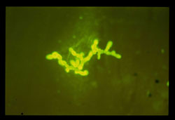 |
Fungal elements observed in abscess aspirate stained by Fungi Fluor (FF) x400 are septate in nature. Fluorescent stain is not suitable to show pigment in the fungal cell wall. The smear was observed under the bright field by switching off the UV filter and turning on white light. | ||||
Figure 2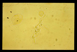 |
The structures observed in wet preparation x400 under the bright field showing fungal elements as pale and not hyaline in nature. | ||||
Figure 3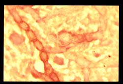 |
Gram smear x1000 of abscess aspirate showing oval cells in chains. Pseudohyphae looking structures often found in clinical specimens containing dark fungi (personal experience). Cell wall of fungal elements is some what prominent and darker. However, not all dark fungi present in the specimen display dark cell wall. | ||||
Figure 4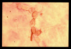 |
Dark fungi suspected by routine staining procedures must be confirmed by special melanin stain known as Fontana Masson (FM). In the image FM x1000 morphology of the fungal elements remains similar to Gram stain. The cell wall is darker, confirms dematiaceous fungus involved. | ||||
Not all fungi showing pale darker cell wall in Wet Prep or Gram stain are true dematiaceous. Figs. 5 & 6 show structures that mimic darker cell wall and Fig. 7 is GMS x1000 showing darkness of septate hyphae (fungal elements). This darkness does not confirm the presence of melanin pigment since the end point of GMS is to stain all fungi as dark. This fact is proven in this example that Fig. 8 FM stain x1000 is negative for melanin pigment. The fungus in question is not dark in nature. This fungus was identified as Aspergillus terreus that belongs to hyaline group. |
|||||
Figure 5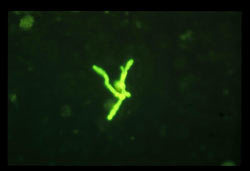 |
Figure 6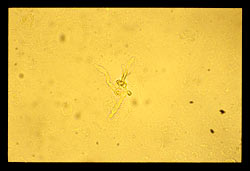 |
||||
Figure 7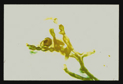 |
Figure 8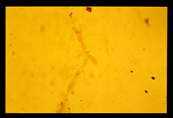 |
||||
Some dark fungi do not display dark cell wall in Gram stained smear. Such fungi may be suspected as dark by morphology and confirmed by melanin stain. Fig. 9 Gram x1000 & Fig. 10 FF x400 do not show pigmented cell wall. However, morphology of the filamentous form appears to be different and not fitting into pseudohyphae or hyaline septate hyphae categories. Fig. 11 stained by FM x1000 showing brown pigmentation in the cell wall. |
|||||
Figure 9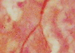 |
Figure 10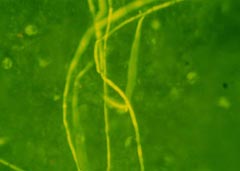 |
||||
Figure 11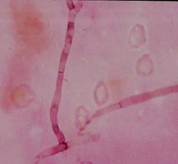 |
|||||
| Gallery Menu :|: Page 1 :|: Page 2 :|: Page 3 :|: Page 4 |
© Copyright 1999-2007 Department of Microbiology, Mount Sinai Hospital, Toronto, Canada. All rights reserved.
|