|
||||||

|
||||||||||||||||||||
|
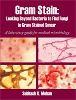 Subhash Mohan has published a book on
“Gram Stain: Looking Beyond Bacteria to Find Fungi in Gram Stained Smear”
(A Laboratory Guide for Medical Microbiology). This book is based on experience containing over 340 colored microscopic images.
It contains flow charts, methods and many cases in all groups of fungi describing decision making insight.
This book also has a quiz section for practice followed by the answers.
Subhash Mohan has published a book on
“Gram Stain: Looking Beyond Bacteria to Find Fungi in Gram Stained Smear”
(A Laboratory Guide for Medical Microbiology). This book is based on experience containing over 340 colored microscopic images.
It contains flow charts, methods and many cases in all groups of fungi describing decision making insight.
This book also has a quiz section for practice followed by the answers.
Routine microbiology receives clinical specimens for C & S. Many of these specimens require direct smear examination a simple staining procedure such as Gram stain. The routine technologist in microbiology examining Gram smears usually search for bacteria and yeasts in direct smears and may miss the presence of fungi. Fungi present in direct Gram smears may remain undetected due to the lack of experience in mycology. General microbiology staff needs to be aware of and learn to recognize objects other than bacteria in direct gram smears.
|
|||||
| Candida albicans | Candida albicans | Candida albicans | |||
| Sporothrix schenckii | Exophiala jeanselmei | Histoplasma capsulatum | |||
| Histoplasma capsulatum | Histoplasma capsulatum | Torulopsis glabrata | |||
| Malassezia furfur | Coccidioides immitis | Coccidioides immitis | |||
| Aspergillus flavus | Aspergillus flavus | Aspergillus fumigatus | |||
| Aspergillus fumigatus | Aspergillus terreus | Aspergillus fumigatus | |||
| Aspergillus niger | Aspergillus fumigatus | Paecilomyces lilacinus | |||
| Ramichloridium mackenziei | Ramichloridium mackenziei | Ramichloridium mackenziei | |||
| Fusarium species | Cryptococcus neoformans | Cryptococcus neoformans | |||
| Rhizopus species | Rhizopus species | Rhizopus species | |||
| Blastomyces dermatitidis | Blastomyces dermatitidis | Rhinosporidium seeberi | |||
| Rhinosporidium seeberi | Penicillium marneffei | Penicillium marneffei | |||
| Penicillium species | Acanthamoeba cysts | Acanthamoeba cysts | |||
| Microsporidia | Microsporidia | Microsporidia | |||
| Prototheca wickerhamii | Prototheca wickerhamii | Prototheca wickerhamii | |||
© Copyright 1999-2007 Department of Microbiology, Mount Sinai Hospital, Toronto, Canada. All rights reserved.
|